DiatomBase image

Pseudo-nitzschia abrensis
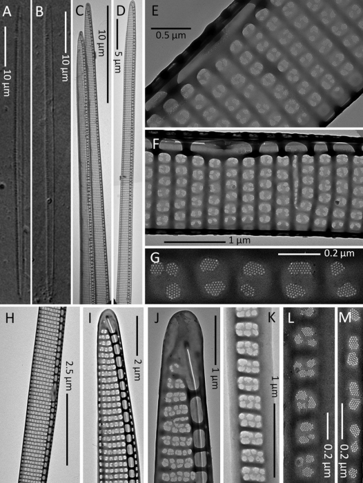
Description (A and B) Light micrographs.(C–M) TEM micrographs.
(A and B) Whole valves.
(C and D) Part of the cell in valve view.
(E) Detail of the central part of a valve showing the central nodule and the striae with a row of poroids split into two sectors (seldom three or four).
(F) Part of a valve showing the central nodule and the poroid arrangement.
(G) Detail of the poroid structure.
(H) Central part of a valve.
(I) Apical part of a valve showing the proximal and distal mantles.
(J) Valve end. (K) Valvocopula.
(L and M) Different cingular bands.
(A–D, G–L) Strain Ner-J2.
(E, F, and M) Strain Ner-J3. ·
 Comment (0)
Disclaimer: DiatomBase does not exercise any editorial control over the information displayed here. However, if you come across any misidentifications, spelling mistakes or low quality pictures, your comments would be very much appreciated. You can reach us by emailing info@marinespecies.org, we will correct the information or remove the image from the website when necessary or in case of doubt.